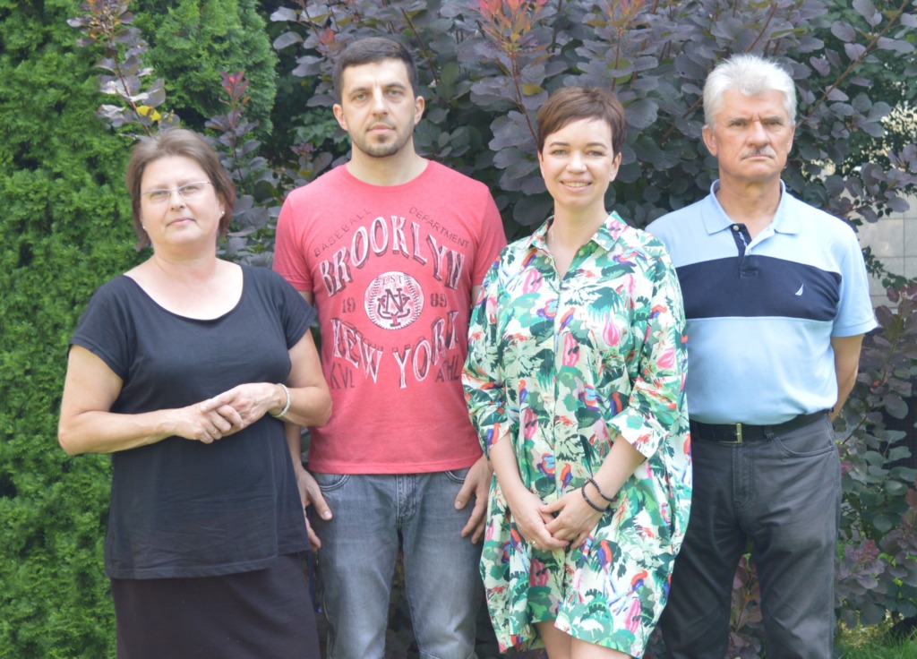
- Head of laboratory
- Scientific Staff
- Technician and administration staff
- Laboratory of Electron Microscopy is a part of the Neurobiology Center of the Nencki Institute of Experimental Biology PAS
- Histology Unit
- Selected publications
Head of laboratory

Laboratory of Electron Microscopy is a part of the Neurobiology Center of the Nencki Institute of Experimental Biology PAS
The Laboratory is equipped with:
– transmission electron microscope JEM 1400 (JEOL Co., Japan, 2008) with energy-dispersive full range X-ray microanalysis system (EDS INCA Energy TEM, Oxford Instruments, UK), tomographic holder and 11 Megapixel TEM Camera MORADA G2 (EMSIS GmbH, Germany);
– transmission electron microscope JEM 1200EX (JEOL Co., Japan 1986);
– ultramicrotomes (LKB, Sweden and Leica, Austria);
– specimen trimming device EM TRIM2 (Leica, Austria);
– critical point drying system (Polaron, UK);
– vacuum evaporator (JEOL Co., Japan);
– EM PACT2 with AFS2 equipment for high-pressure freezing (HPF) and automatic freeze substitution (AFS) (Leica, Austria).
We provide expertise and assistance in electron microscopic studies:
– qualitative and quantitative X-ray microanalysis and elements mapping (EDS – Energy Dispersive Spectroscopy);
– 3D imaging as well as reconstruction and ultrastructural analysis of biological samples;
– imaging of macromolecular assemblies of proteins;
– sample (cells and tissues) preparation for TEM analysis with chemical fixation and embedding procedures with epoxy resins (Durcupan, Epon) or physical immobilization using high-pressure freezing and freeze substitution (Lowicryl);
– sectioning with ultramicrotome for TEM analysis;
– mild drying of the biological samples in liquid CO2 (CPD – critical point drying);
– coating of specimen grids with carbon for TEM analysis;
– coating of the biological samples with neutral metals for SEM analysis.
Annually, ~20 000 digital electron micrographs and ~ 600 EDS spectra are collected in our core facility. We are also involved in education and dissemination activities: lectures, workshops and practical training.
We are open to scientific cooperation.
For reservation please use the link: http://intra.nencki.gov.pl/category/kalendarze/
External users can reserve services via the e-mail: tem@nencki.gov.pl or phone: (48 22) 58 92 374.
Histology Unit
Histology Unit delivers a high standard of service in histological techniques. The Unit also provides access to the following equipments:
– cryostat Leica CM 1950;
– cryostat Microm HM 550;
– sliding microtome HP 440 E.
We provide expertise and assistance in:
– preparation of sample for sectioning on cryostat different animal tissues (freezing method);
– preparation of sample for sectioning on microtome different animal tissues (paraffin method);
– histological staining: H&E, Nissl, Klüver, Timm, CO, AChE;
– other staining methods.
We also provide training in sectioning on cryostats and microtomes, paraffin and celloidin embedding methods, histological staining.
For any additional information or enquiries please contact our histology specialist Agnieszka Kępczyńska, e-mail: histology.unit@nencki.gov.pl
For reservation please use the link: http://intra.nencki.gov.pl/category/kalendarze/
External users can reserve this service via e-mail: histology.unit@nencki.gov.pl or phone: (48 22) 58 92396.
Selected publications
Nieznanska H., Bandyszewska M., Surewicz K., Zajkowski T., Surewicz W.K., Nieznanski K. (2018) Identification of prion protein-derived peptides of potential use in Alzheimer’s disease therapy. Biochim. Biophys. Acta, 1864, 2143-2153.
Strzelecka-Kiliszek A., Bozycki L., Mebarekb S., Buchetb R., Pikula S. (2017) Characteristics of minerals in vesicles produced by human osteoblasts hFOB 1.19 and osteosarcoma Saos-2 cells stimulated for mineralization. Journal of Inorganic. Biochemistry, 171, 100–107.
Waclawek E., Joachimiak E., Hall MH., Fabczak H., Wloga D. (2017) Regulation of katanin activity in the ciliate Tetrahymena thermophila. Mol. Microbiol., 103,134-150.
Bilska-Kos A., Solecka D., Dziewulska A., Ochodzki P., Jończyk M., Bilski H., Sowiński P. (2017) Low temperature caused modifications in the arrangement of cell wall pectins due to changes of osmotic potential of cells of maize leaves (Zea mays L.). Protoplasma, 254, 713-724.
Zajkowski T., Nieznanska H., Nieznanski K. (2015) Stabilization of microtubular cytoskeleton protects neurons from toxicity of N-terminal fragment of cytosolic prion protein. Biochim. Biophys. Acta, 1853, 2228–2239.

