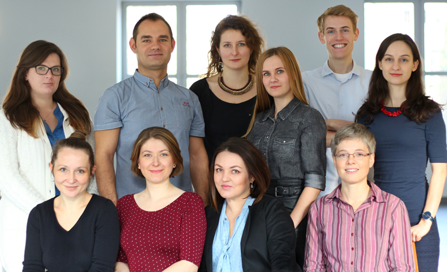
- Head of laboratory
- Scientific Staff
- Technician and administration staff
- PhD Students
- About Laboratory of Cytometry
- Core-facility activities
- Equipment
- Methods/Applications in flow cytometry
- Research - currently we investigate
- Service
- Selected publications
Head of laboratory

About Laboratory of Cytometry
The Laboratory of Cytometry has been established in June 2010 to provide high quality expertise, equipment and multicolor flow cytometry service. Our vision is to offer service and scientific collaboration to meet researchers’ specific needs by dedicated help with design of experiments, acquisition and advanced data analysis.
Our research is dedicated to cancer biology. The goal is to understand mechanisms of disease progression and development of resistance and discover potential molecular targets in leukemia to develop and propose potential therapies, with special interest in intercellular interactions and the role of leukemia microenvironment.
More information about laboratory on the web page: http://piwocka-lab.nencki.edu.
Core-facility activities
1. The Laboratory provides the core-facility service for investigators from the Nencki Institute and other scientific and R&D institutions;
2. We offer expertise and technical assistance in flow cytometry and cell sorting. We provide the expert consultation for experiment design, fluorochrome selection and data analysis;
3. The Laboratory is involved in the basic research and innovative projects;
4. Education – we organize lectures, training courses and hands-on workshops for beginners and advanced researchers.
Equipment
BD FACSAria II cell sorter – equipped in the 405 nm, 488 nm and 635 nm lasers
Description:
BD FACSAria II is a high-speed cell sorter. It is equipped with a fixed-alignment flow cell cuvette which provides superior fluorescence sensitivity. It can simultaneously detect up to 11 parameters (2 scatter signals and up to 9 fluorescent parameters) by utilizing 3 air-cooled, solid state lasers (laser outputs at 405nm, 488nm and 633nm) and a fixed optical alignment system allowing for multiple configurations and a wide range of fluorochromes. The FACSAria allows for digital acquisition rates of up to 70,000 cells per second. Sorting into different types of test tubes (microtubes, 5 and 15 ml tubes) is possible. The cytometer can sort using Automated Cell Deposition Unit (ACDU) to 96-well plates or on the microscope slide. The sorter is equipped with an Aerosol Management System that evacuates any aerosols from the sort chamber and has a refrigeration system that allows sorting the sample in 4, 21 or 37 degrees.
Accessories: BD FACSDiva software
Cytek Aurora spectral cytometer – equipped in the 355 nm, 405 nm, 488 nm, 561 nm, 640 nm lasers
Description:
With five lasers, three scattering channels (FSC, blue laser SSC and violet laser SSC) and 64 fluorescence channels, the Aurora suits every laboratory’s needs, from simple to high complexity applications. The detection of some fluorochrome combinations by conventional flow cytometry presents a challenge due to high amounts of spectral overlap. The Aurora addresses this challenge by using differences in full emission spectra signatures across all lasers to clearly resolve these combinations, even if the populations are co-expressed.
The state-of-the-art optics and low-noise electronics provide excellent sensitivity and resolution. Flat-top laser beam profiles, combined with a uniquely designed fluidics system, translate to outstanding performance at high sample flow rates.
This system delivers high quality data where rare and dim populations are easily resolved, regardless of assay complexity.
Accessories: SpectroFlo® software
BD LSR Fortessa Analyser – equipped in the 355 nm, 405 nm, 488 nm and 635 nm lasers
The BD LSRFortessa™ cell analyzer the laboratory is equipped with offers the ultimate in choice for flow cytometry, providing performance and consistency. The system includes a novel collection optics that reduce excitation losses and improve light collection efficiency. The result is optical efficiency that delivers maximal sensitivity and resolution for multicolour applications. An affordable choice to fit most flow cytometry analyzer needs, the BD LSRFortessa™ system has been configured with 4 lasers — blue (488 nm), red (640 nm), violet (405 nm) and UV (355 nm). This enables the detection of up to 13 colors simultaneously. The high throughput sampler (HTS) allows cells to be taken from a 96 or 384 well plate for screening assays.
Accessories: BD FACSDiva software
Cytometer BD FACSCalibur – equipped in the 488 nm laser and red diode 635 nm
Description:
The laboratory possesses two FACSCaliburs, which are benchtop flow cytometers that provide standard multicolor analysis. They are equipped with two lasers, an air-cooled argon laser (488nm) and a red diode laser (635nm), which are spatially separated for high sensitivity needed for multicolor analysis. Up to four colors can be analyzed on this system using the 488 and 635 lasers. The detectors available from the 488 laser are 530/30, 585/42, >670, and from the 635 laser, 661/16. The BD FACSCalibur flow cytometer provides flexibility to support a wide variety of research such as cell analysis, assay development, verification, and isolation of cellular populations of interest.
Capillary cytometer Merck Millipore Guava easyCyte – equipped in the 488 nm and 640 nm
Description:
Microcapillary Guava easyCyte8HT system is very simple to operate and an alternative technology in flow cytometry. It is equipped with solid state 488nm and 640nm lasers, allowing simultaneous detection for up to 6 fluorescent parameters. The cytometer utilizes small sample volumes and requires no sheath fluid to minimize waste. The cytometer possesses a robotic sample tray that automatically handles a 96-well plate. The powerful InCyte software has a drop & drag menu, HeatMap for data presentation. It enables semi- automated compensation with ability to post-acquisition compensation and automated calculation of IC50/EC50 curves.
Accessories: InCyte software
Methods/Applications in flow cytometry
– multicolor/multiparameter cytometry (panel designing)
– cell sorting
– immunophenotyping
– analysis of cell survival and various parameters of apoptosis
– PrimeFlow – detection of RNA by cytometry
– DNA damage analysis
– analysis of cell cycle and proliferation
– measurement of mitochondrial membrane potential
– cytometric analysis of signaling pathways
– immunocytochemical staining of intracellular and membrane proteins
– measurement of ROS production
– determination of cytokines
– cytometry based FRET- assessment of protein-protein interaction using fluorescent tags
In our research we apply a broad spectrum of cellular and molecular biology as well as bioimaging methods
Research - currently we investigate
– mechanisms promoting leukemia progression and therapy resistance in leukemia; looking for potential therapeutic targets and strategies
– integrated Stress Response pathways in leukemia
– non-classical mechanisms of BRCA1/2 deficiencies in leukemia and sensitivity to personalized therapy by PARP inhibitors
– the leukemia-bone marrow stroma interactions
– direct intercellular connections within leukemia microenvironment by tunneling nanotubes (TNTs)
– leukemic extracellular vesicles and influence on immunosuppression
– protein markers for BRCA1/2 deficiency and PARP1 inhibitors in cancer
Service
We provide service for all business entities, including research institutions, as well as commercial and R&D institutions.
We offer:
– Planning and performance of experiments in a range of our cytometric expertize
– Data analysis, visualization and interpretation (FACSDiva, FloJo, FlowSOM, tSNE)
– Consultations in experiments planning, designing, data analysis and interpretation
– Trainings and workshops
Fees and detailed information about the provided service on the web page: https://piwocka-lab.nencki.edu.pl/home-page/core-facility-service
The service is co-financed by the TEAM-TECH Core Facility Plus/2017-2/2 (POIR.04.04.00-00-23C2/17) grant – FLOW-PROSPER

Selected publications
Szymańska E, Nowak P, Kolmus K, Cybulska M, Goryca K, Derezińska-Wołek E, Szumera-Ciećkiewicz A, Brewińska-Olchowik M, Grochowska A, Piwocka K, Prochorec-Sobieszek M, Mikula M, Miączyńska M. Synthetic lethality between VPS4A and VPS4B triggers an inflammatory response in colorectal cancer. EMBO Mol Med. 2020 Jan 13:e10812.
Swatler J, Dudka W, Bugajski L, Brewinska-Olchowik M, Kozlowska E, Piwocka K. Chronic myeloid leukemia-derived extracellular vesicles increase Foxp3 level and suppressive activity of thymic regulatory T cells. Eur J Immunol. 2019 Nov 23.
Kolba MD, Dudka W, Zaręba-Kozioł M, Kominek A, Ronchi P, Turos L, Chroscicki P, Wlodarczyk J, Schwab Y, Klejman A, Cysewski D, Srpan K, Davis DM, Piwocka K. Tunneling nanotube-mediated intercellular vesicle and protein transfer in the stroma-provided imatinib resistance in chronic myeloid leukemia cells. Cell Death Dis. 2019 Oct 28;10(11):817.
Podszywalow-Bartnicka P, Maifrede S, Le BV, Nieborowska-Skorska M, Piwocka K, Skorski T. PARP1 inhibitor eliminated imatinib-refractory chronic myeloid leukemia cells in bone marrow microenvironment conditions. Leuk Lymphoma. 2019 Jan;60(1):262-264.
Maifrede S, Nieborowska-Skorska M, Sullivan-Reed K, Dasgupta Y, Podszywalow-Bartnicka P, Le BV, Solecka M, Lian Z, Belyaeva EA, Nersesyan A, Machnicki MM, Toma M, Chatain N, Rydzanicz M, Zhao H, Jelinek J, Piwocka K, Sliwinski T, Stoklosa T, Ploski R, Fischer T, Sykes SM, Koschmieder S, Bullinger L, Valent P, Wasik MA, Huang J, Skorski T. Tyrosine kinase inhibitor-induced defects in DNA repair sensitize FLT3(ITD)-positive leukemia cells to PARP1 inhibitors. Blood. 2018 Jul 5;132(1):67-77.
Nieborowska-Skorska M, Sullivan K, Dasgupta Y, Podszywalow-Bartnicka P, Hoser G, Maifrede S, Martinez E, Di Marcantonio D, Bolton-Gillespie E, Cramer-Morales K, Lee J, Li M, Slupianek A, Gritsyuk D, Cerny-Reiterer S, Seferynska I, Stoklosa T, Bullinger L, Zhao H, Gorbunova V, Piwocka K, Valent P, Civin CI, Muschen M, Dick JE, Wang JC, Bhatia S, Bhatia R, Eppert K, Minden MD, Sykes SM, Skorski T. 18. Gene expression and mutation-guided synthetic lethality eradicates proliferating and quiescent leukemia cells. J Clin Invest. 2017 Jun 1;127(6):2392-2406.
Wolczyk M, Podszywalow-Bartnicka P, Bugajski L, Piwocka K. Stress granules assembly affects detection of mRNA in living cells by the NanoFlares; an important aspect of the technology. Biochim Biophys Acta Gen Subj. 2017 May;1861(5 Pt A):1024-1035.

